CAP Inspection Readiness.
Stress Reduced.
Discrepancies Minimized.
Custom compliance binders, SOP optimization, and inspection readiness support for Anatomic Pathology labs preparing for CAP inspections.
How Ion Helps Your Laboratory Prepare
Preparing for inspection can feel overwhelming, especially in labs where the supervisors are also responsible for daily bench work.
Ion provides a structured approach to readiness by organizing documentation, identifying compliance gaps, and building systems that allow inspectors to easily verify laboratory practices.
The Ion Method
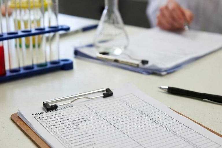
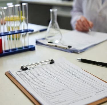
How Ion helps histology labs prepare for inspection.
Learn the Lab
Understand the laboratory workflow, documentation practices, and inspection timeline.
\/
Assess Documentation
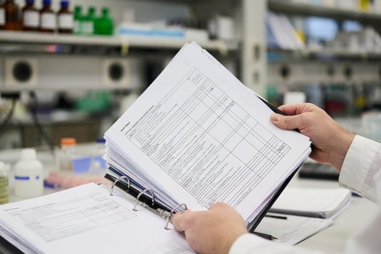
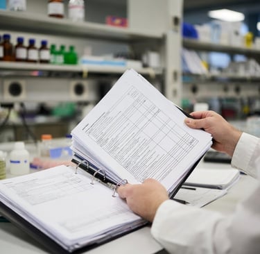
Review SOPs, QC records, and existing compliance documentation.
\/
Identify Gaps
Create a CAP Readiness Gap Assessment Report with prioritized actions.
\/
Map the Evidence
Link the CAP checklist requirements to documentation using the Ion Crosswalk Matrix.
\/
Build the Inspection Binder
Organize documentation so inspectors can easily verify compliance.
\/
Inspection Day Becomes Routine
Staff remain calm, documentation is easy to retrieve, and inspections run smoothly.
Who Ion Helps
Ion supports Anatomic Pathology laboratories preparing for CAP or CLIA inspections, particularly labs where the supervisor balances compliance responsibilities with daily technical work.
Many laboratories do not have dedicated compliance staff, leaving the supervisor responsible for both operational workflow and inspection readiness.
Ion steps in to handle the compliance groundwork, reviewing documentation, identifying gaps, and building organized inspection systems, so laboratory leaders can remain focused on running their lab and supporting patient care.
Ion is especially helpful for:
> Independent pathology laboratories
> Physician-owned laboratories
> Hospital histology labs with limited compliance support
> New laboratories preparing for their first inspection
> Labs that have struggled with inspection preparation in the past
> Supervisors who are responsible for both bench work and compliance
Services
Ion provides practical compliance support that helps histology laboratories prepare for inspection without overwhelming supervisors responsible for daily lab operations.
Each engagement is tailored to the laboratory's needs, with Ion handling the detailed documentation work required to demonstrate inspection readiness.
Laboratories may engage Ion for a single service or for comprehensive inspection preparation support, depending on their needs.
CAP Readiness Gap Assessment
A structured review of laboratory documentation, SOPs, and quality records to identify compliance gaps and prioritize preparation efforts before inspection.
Inspection Documentation Crosswalk
Mapping CAP checklist requirements to supporting documentation so inspectors can easily verify compliance.
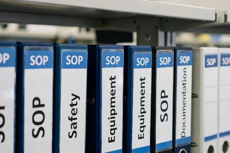
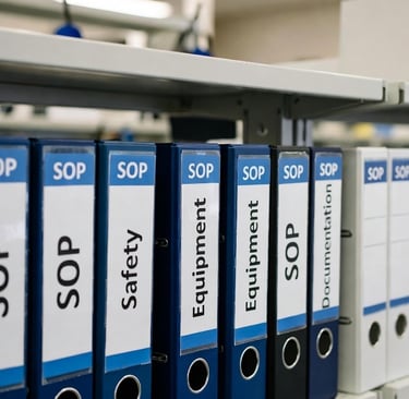
Inspection Binder Development
Creation of organized documentation systems that allow inspectors to quickly locate required materials during inspection.
Inspection Preparation Support
Hands-on assistance organizing documentation and preparing laboratory systems as inspection approaches.
Request a Consultation
If you are preparing for an upcoming inspection or would like support improving documentation systems in your laboratory, Ion Histology offers practical, structured consulting tailored to your needs.
Initial consultations are designed to understand your laboratory's current processes, identify areas of friction, and determine if Ion's services are the right fit.
There is no obligation to proceed following an initial consultation.